Human papillomavirus type 16 Recombinants

Abstract

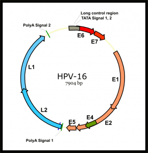
Intratypic diversity of the human papillomavirus (HPV) type 16 Recombinants genome is generally characterized by a point mutation, insertion, and/or deletion. Using PCR-based cloning and sequencing, we detected concurrent infection with 8 HPV16 variants in a woman enrolled in the ASCUS-LSIL classification study. The European variant was the main variant; each of the 7 minor variants had partial DNA sequences identical to the European variant and another part identical to the African variant 2. At a follow-up visit, only one African variant 2 of HPV16 was detected. The results of the present study suggest the presence of intratympanic recombination of the HPV genome in natural infection.

Bacterial culture strains and conditions.

The strain used in this study was L. casei CECT 5275 [=ATCC 393(pLZ15-)]. Wild-type L. casei was grown in MRS medium (Difco) at 37°C without shaking. For expression analysis, recombinant L. casei was cultured in basal MRS medium (10 g peptone per litre, 8 g meat extract per litre, 4 g yeast extract per litre, 2 g monobasic potassium phosphate per litre). litre, 5 g sodium acetate per litre, 2 g diammonium citrate per litre, 0.2 g magnesium sulfate per litre, 0.03 g manganese sulfate per litre, 1 mL Tween 80 per litre, buffered with 0.2 M potassium phosphate [pH 7.0]) supplemented with 0.5% lactose as a carbon source for induction and with 0.5% glucose as a carbon source for repression of the operon promoter. lactose from L. casei. Escherichia coli DH5α was grown in LB medium at 37°C with shaking for replication of expression vectors in cloning procedures. The erythromycin concentrations used for the selection of recombinant strains were 350 μg/ml and 5 μg/ml for E. coli and L. casei, respectively.

Electron microscope.

L. casei and L. casei/L1 were grown until the OD600 was 2. Cultures (2 ml) were centrifuged at 5000 × g for 10 min and each pellet was washed three times in saline. Bacteria were fixed with 3% paraformaldehyde (Sigma) for 1 hour at room temperature. Appropriate volumes of fixed intact bacteria were adhered to carbon-coated grids for 1 min and air-dried. Some samples were negatively stained with 2% uranyl acetate for 1 min. Samples were examined with a Zeiss EM 109 transmission electron microscope operated at 80 kV.

Immunofluorescence.

L. casei and L. casei/L1 were washed and fixed as described above. Cells then adhered to polylysine-coated microscopic glass slides. To detect VLPs produced by L. casei by immunofluorescence, slides were incubated with an anti-HPV-16 VLP conformational antibody, H16.V5 (10) (kindly provided by N. Christensen, Department of Microbiology and Immunology, Pennsylvania State University College of Medicine, Hershey, PA), diluted in PBS containing 0.01% Tween 20 and 0.5% BSA for 2 h at 37 °C.

Cells were then washed five times in PBS for 5 minutes and incubated with fluorescein isothiocyanate (FITC)-conjugated anti-mouse IgG (Sigma) diluted in PBS containing 0.01% Tween 20 and 1% BSA. 5% for 1 hour at 37°C. After five washes in PBS, slides were air-dried, embedded in Mowiol (Calbiochem), covered with coverslips, and stored at 4°C until use. Fluorescence was visualized by exposing slides to a filter with an excitation wavelength of 488 nm from a Zeiss LSM 510 META confocal microscope.

Immunization of mice.

Six to eight-week-old female BALB/c mice from the Butantan Institute were used for subcutaneous immunization experiments. L. casei and L. casei/L1 were cultured until the OD600 was 2. The bacteria were collected by centrifugation at 5000 × g and washed three times with non-pyrogenic saline, and then the concentration was adjusted to 109 cells per 100 μl.

Groups of five mice were inoculated with 100 µl of saline, L. casei or L. casei/L1 on two consecutive days. Three administrations, one sensitizing and two boosting, were performed at 2-week intervals, for a total of six administrations. Ten days after the last administration, animals were bled through the retroorbital plexus and pooled sera were collected and stored at -20°C until use. The results described below are representative of two experiments.

ELISA for the detection of anti-HPV-16 VLP antibodies.

ELISA plates were coated with 100 ng of VLPs derived from S. frugiperda insect cells diluted in PBS (assembled VLPs) or 0.2 M carbonate buffer (pH 9.6) (disassembled VLPs) (23) at 4 °C for 16 h. Pooled sera were tested for the presence of anti-L1 IgG as previously described (1). The titer was defined as the dilution at which the absorbance at 492nm was 0.15. The results presented below are the means of two independent immunization experiments.

Escherichia coli O6:H1 Recombinants

Species

Escherichia coli O6:H1 recombinant (strain CFT073 / ATCC 700928 / UPEC)

Fountain: E. coli

Label information: N-terminal 10xHis-SUMO-tagged and C-terminal Myc-tagged

Estimated response time: 7-11 business days

Protein type: recombinant protein

Gene name: ppiA

Alternative names

cyclophilin A; Rotamase A

Uniprot: P0AFL4

Expression Region: 25-190yy

Sequence information: Total length of the mature protein

Theoretical MW: 38.1 kDa

Purity: More than 85% as determined by SDS-PAGE.

Storage buffer

If the dosage form is liquid, the default storage buffer is a Tris/PBS based buffer, 5%-50% glycerol. If the administration form is a lyophilized powder, the buffer before lyophilization is Tris/PBS-based buffer, 6% trehalose, pH 8.0.

Endotoxin Level: Not tested

Shipping condition: ice packs

Storage

Short term: -20°C; Long life: -80°C. Minimize freeze-thaw cycles.

Date of Expiry: 1 year

Investigation area: Immunology

Restriction

For research use only. Not for use in diagnostic procedures, drug use, or for administration to humans or animals.

Relevance

PIases accelerate protein folding. It catalyzes the cis-trans isomerization of proline imidic peptide bonds in oligopeptides.

Function

PIases accelerate protein folding. It catalyzes the cis-trans isomerization of proline imidic peptide bonds in oligopeptides (By similarity).

Involvement in the disease

PIases accelerate protein folding. It catalyzes the cis-trans isomerization of proline imidic peptide bonds in oligopeptides (By similarity).

Salmonella typhi Recombinants

Abstract

Typhoid fever caused by Salmonella typhi recombinant is an endemic disease in Asia, including Indonesia. According to data from the World Health Organization (WHO), there are 81% of every 100,000 people in Indonesia infected with typhoid fever. Based on this, the development of a typhoid fever detection kit that is simple, rapid, specific and sensitive is still needed. One of the currently available detection kit developments is the antigen-antibody interaction approach.

This study aims to obtain complete information on the purification of recombinant Fim-C-S. typhi proteins as antigens for raw materials for the manufacture of typhoid fever detection kits. This study uses the IMAC method with Ni2+ metal, which has advantages in high selectivity and purity. The results of this study report the purification of recombinant Fim-C-S. Typhi protein inclusion bodies with variations in the 3-, 5-, and 7-fold binding and washing process gave corresponding percentage yields of 37.84, 39.44, and 38.21% pure protein.

Previous studies also reported variations in binding and washout at 2-, 4-, and 6-fold and gave percentage yields of 35.37, 39.11, and 40.49%, respectively. Based on the data, we conclude that the 6-fold variation is the best repeat variation to obtain the highest percentage yield so that it can be used as a reference in large-scale purification.

Product name

Salmonella typhi uspA recombinant protein (His tag)

Purity: > 90% SDS-PAGE.

Expression system: Mammal

Accession: Q8Z268

Protein length: full-length protein

Animal-free: No

Nature: recombinant

Species: Salmonella typhi

Amino acids: 2 to 144

Tags: Your N-Terminus Tag

Additional sequence information: mature chain

Specifications

Our Abpromise warranty covers the use of ab267923 in the following tested applications. Application notes include recommended starting dilutions; the end-user must determine the optimal dilutions/concentrations.

Applications: SDS PAGE

Form: Liquid

Concentration: 10 µg to 0.21 mg/mL

Stability and Storage

Shipped at 4°C. Store at +4°C for the short term (1-2 weeks). At the time of aliquot delivery. Store at -20°C or -80°C. Avoid freeze/thaw cycle.

  • pH: 7.2
  • Components: PBS, 6% trehalose

Lyophilized from.

T-SPOT.TB

Background:

The diagnosis of tuberculosis (TB) remains difficult. Previous reports have shown that the T-SPOT.TB assay may be a more promising diagnostic tool for TB; however, further study is needed to assess the diagnostic value of T-SPOT.TB for specific populations in a high-prevalence setting.

Methods:

In this present study, we conducted stratified and comparable analyzes to explore the clinical value and limitations of the T-SPOT.TB assay in TB diagnosis in a high-prevalence TB setting in southern China. A total of 413 subjects were included in the study, including 163 with pulmonary tuberculosis (PTB), 39 with extrapulmonary tuberculosis (EPTB), 106 with non-tuberculosis pulmonary diseases (NTBPD), 20 medical staff, and 85 healthy controls.

  • Subjects

In total, 20,332 samples were collected from patients with suspected TB. The average age of the participants was 53.15 ± 18.16 years, and they were admitted to the First Affiliated Hospital of Xi’an Jiaotong University from July 2013 to May 2017. Specifically, there were 11,453 males between 6 months and 96 years of age ( mean age: 54.70 ± 18.45), and 8,879 women between 3 months and 96 years (mean age: 52.53 ± 17.71 years). All patients were enrolled according to the following criteria: 1) subjects did not have heart, liver, or kidney disease and did not have HIV infection; 2) patients were suspected of having TB; 3) The patients were not taking any therapy involving immunosuppression or potentiating medication. Exclusion criteria included: 1) cases lost to follow-up; 2) cases lost due to death; 3) cases that were not diagnosed as TB at endpoints.

Informed consent was obtained from all participants (informed consent of patients under 16 years of age was obtained from their guardians), and this study was approved by the ethics committee of the First Affiliated Hospital of Xi’an Jiaotong University (NO .XJTU1AF2018LSK-161).

  • Diagnostic criteria for different groups.

In light of the diagnostic criteria for pulmonary TB established by the Ministry of Health of the People’s Republic of China, the patients included were grouped as follows: 1) LTBI: the tuberculin test was positive and patients who had no history of the Bacillus Calmette – Guerin (BCG) vaccination; or T-SPOT.TB test result was positive and there was no clinical manifestation of TB and corresponding evidence of aetiology, pathology, and imaging. 2) Active TB: This required that tubercle bacilli be detected from bacterial cultures or sputum smears, that caseate or giant Langhans cells be seen by pathological examination, and that anti-TB therapy is effective with relevant imaging support.

In addition, active TB was divided into three subgroups based on pathological site: pulmonary TB (PTB), pulmonary and extrapulmonary TB (PETB), and extrapulmonary TB (EPTB). 3) Old TB: this required that subjects with a history of TB were cured, but pathological changes were found according to imaging diagnosis, that no symptoms of TB intoxication were observed and the aetiology and pathology tests were negative, and that patients with these characteristics were diagnosed with “other diseases”.

  • Sample collection and handling

Peripheral venous blood samples of 5 ml were obtained from patients with suspected TB using lithium heparin anticoagulant tubes. Mononuclear cells were then isolated to prepare a cell suspension. Finally, the T-SPOT.TB assay (Oxford Immunotec, Ltd., Abingdon, UK) was performed as follows: Briefly, the cell suspension was plated onto T-SPOT.TB plates and incubated with ESAT-6 ( specific antigen), CFP-10 (specific antigen), positive control or negative control, respectively. Next, 100 μl of cell suspension were added to the corresponding microwells and cultured in an incubator with 5% CO2 at 37  °C.

The microwells were then washed four times with phosphate-buffered solution (PBS) before 50μl of secondary antibody solution was added to each well, and the assay was incubated for 1h at 2–8°C. Subsequently, 50 μl of the chromogenic agent were added and the plate was processed avoiding light for 7 to 12 min before finishing with distilled water. The number of dots was measured, where one dot represented a T cell that could secrete specific cytokines. The final interpretation of the results was according to the following criteria: 1) The results were considered positive in two scenarios: first if the number of spots in the negative control group was less than 6, and the number of spots in CFP -10 or ESAT-6 wells was 6 points higher than that of the negative control.

Second, if the spot count in the negative control group was 6 to 10, and the number of CFP-10 or ESAT-6 spots was more than double that of the negative control. 2) Results were considered negative if the spot number did not meet the above criteria and the positive control performed normally. Finally, we performed statistical analyzes of the number of points and the points represented the proportion of the total points (composition rate) in different types of diseases (LTBI, active TB and old TB) and different subgroups of active TB (PTB, PETB and EPTB). )

Results:

According to T-SPOT.TB, there was a high incidence of latent TB infection (LTBI) in the general population in southern China, especially in the NTBPDS and medical staff. The T-SPOT.TB had a high yield in the diagnosis of active TB (ATB) in a population with a lower risk of TB infection such as the general population, however, the T-SPOT.TB for the diagnosis of ATB in the High-risk population of TB infection populations involving close contacts such as pulmonary disease (PD) patients or medical personnel is unreliable due to LTBI interference.

Under this condition, the value of exclusion from the trial appeared to be better than that of acceptance. We believe that T-SPOT.TB is suitable for screening for both EPTB and ATB combined with diabetes mellitus (DM). However, we found the sensitivity of T-SPOT.TB in the smear-negative population was not as high as in the smear-positive population.

Conclusions:

T-SPOT.TB test results should be interpreted with caution in combination with subject characteristics in a high-prevalence setting.

Keywords:

T-SPOT.TB; diagnostic tool; tuberculosis (TB); Extrapulmonary TB (EPTB)

IMMUNEMED Scrub Typhus Rapid

Rub typhus fast

Orientia tsutsugamushi infection can be diagnosed by detecting Orientia tsutsugamushi-specific IgM and IgG antibodies and screening detection results with superior sensitivity and specificity can be obtained only in 15 minutes.

Very fast

Very quickly interpret the result reading in just 15 minutes.
One step from a physical exam to prescription on the same day.

High accuracy

  • Detection of several serotypes of Orientia tsutsugamushi is possible through the use of special chimeric antigens and supplementary antigens.
  • Higher sensitivity (97.3%, 146/150) and specificity (99.5%, 198/199).1)
  • Diagnose Scrub Typhus with great precision.

Easy and simple to use

  • Use a variety of blood samples, such as whole blood, serum, and plasma.
  • Just 3~4 steps from blood to performance with a very easy and simple process.
  • Optimized in the Point-of-Care diagnosis.

Tsutsugamushi’s disease

Scrub Typhus is an acute febrile illness caused by an infection of Orientia tsutsugamushi. Its clinical symptoms include early fever, chills, and headache that are similar to a cold, and also muscle pain, cough, vomiting, abdominal pain, and throat pain. This disease is transmitted by some species of trombiculid mites (chiggers) and the bite of this mite leaves a characteristic black eschar that is helpful to the physician in making the diagnosis. The condition of a person infected with the disease improves in a day or two if properly treated. However, if it is not correctly, the fever lasts about two weeks. Therefore, an early definitive diagnosis is very important.

Characteristics

  • Qualitative test for the detection of specific IgM and IgG antibodies of Orientia tsutsugamushi existing in human blood (Serum, Plasma and Pureblood).
  • Very easy to diagnose disease progress and reinfection status as IgM and IgG are tested at the same time.
  • Store at room temperature (4–30 °C).
  • Interpret test results very quickly.
  • As a diagnostic kit, it is optimized for on-site diagnosis.

HS-qPCR SYBR Blue (2x)

Characteristics

Enzyme with hot start capability increases the specificity and sensitivity of the reaction, optimized
Activation of DFS-Taq PLUS DNA polymerase requires no more than 5 minutes of heating
High selectivity and reaction yield
The mixture is coloured to facilitate pipetting
Reduced preparation time

Request

  • Real-time PCR with intercalated dye SYBR Green I
  • conventional PCR

Basic Description

BioMaster HS-qPCR SYBR Blue (2×) contains 2× BioMaster HS-qPCR SYBR Blue reaction mix and sterile water. The master mix is ​​developed for quantitative real-time PCR with fluorescent dye SYBR Green I. BioMaster HS-qPCR SYBR Blue (2×) contains all components (except template DNA and primers) required for PCR: HS-qPCR Taq DNA polymerase, dNTP mix, PCR buffer, Mg2+, SYBR Green I and inert dye. The mix is ​​optimized for efficient and reproducible hot-start real-time PCR of genomic, plasmid, and viral DNA samples.

The solution contains substances that increase the half-life and processivity of HS-Taq DNA polymerase by improving its stability during PCR. BioMaster HS-qPCR SYBR Blue (2×) includes components that influence primer annealing temperature and template melting characteristics, allowing you to increase PCR specificity and use templates with complicated spatial structures. The blue hue of the reaction solution provided by the inert dye allows for control when using multi-well plates. The use of the kit saves time and minimizes the risk of contamination due to the reduction in the number of pipetting steps.

Areas of use

Classic PCR with “hot” start

Properties of the reaction mixture

  • optimized for real-time PCR
  • contains substances that allow long-term storage (storing BioMaster HS-qPCR SYBR Blue (2×) at room temperature for one month does not affect PCR efficiency) and several cycles of thawing and freezing

Advantages:

  • Green Sybr mix is coloured for easy pipetting
  • Reduced preparation time
  • Low probability of contamination during the preparation of the PCR solution
  • Standardized conditions of same type reactions (reduction of pipetting error during mixing of PCR components from one experiment to another)
  • Minimized efforts

100 mM UTP

Description

Thermo Scientific UTP (uridine 5′-triphosphate) is supplied as a 100 mM aqueous solution titrated to pH 7.3-7.5 with NaOH. This highly stable nucleotide is greater than 99% pure. You can use UTP in many molecular biology applications.

Reflexes

• Greater than 99% purity confirmed by HPLC
• Functionally tested in in vitro transcription
• High stability: stable for two years at -20°C and after multiple freeze-thaw cycles

Applications

  • aRNA synthesis
  • siRNA synthesis
  • RNA amplification
  • in vitro transcription

For research use only. It should not be used in diagnostic procedures.

Specifications

Conjugate: unconjugated

Concentration: 100mm

Physical Appearance: Colorless transparent liquid

Storage: Store at -20°C. It is recommended to divide the product into small parts after receipt to avoid repeated freezing and thawing.

M.Wt: 550.09 (acid form: 484.09)

Cas No: 63-39-8 (acid form); 19817-92-6 (3Na salt)

Formula: C9H12N2Na3O15P3

Synonyms: Uridine-5′-triphosphate

Chemical name: Sodium ((2R,3S,4R,5R)-5-(2,4-dioxo-3,4-dihydropyrimidin-1(2H)-yl)-3,4-dihydroxytetrahydrofuran-2-yl)methyltriphosphate

Taq DNA Polymerase

General description

FastStart™ Taq DNA Polymerase is a versatile enzyme that can be used in a wide variety of applications and across multiple instrument platforms. This modified thermostable recombinant Taq DNA polymerase is inactive at temperatures below +75°C but is activated by a 2-4 minute heat activation step at +95°C. Because it is inactive at low temperatures, FastStart™ Taq DNA Polymerase cannot elongate non-specific primer-template hybrids that can form at these temperatures. FastStart™ Taq DNA Polymerase is an ideal tool for hot-start PCR because the enzyme remains inactive during PCR setup and before the initial denaturation step.

Request

FastStart™ Taq DNA Polymerase is a thermostable, chemically modified form of recombinant Taq DNA polymerase. The enzyme is inactive at +15 to +25°C during PCR setup and then activated at +95°C during initial denaturation. This enzyme offers superior results due to its unique enzyme design and optimized buffer system. FastStart Taq DNA Polymerase is an ideal tool for hot-start PCR because the enzyme remains inactive during the PCR setup and before the initial denaturation step.

It can be requested for:

  • PCR
  • multiplex PCR
  • Difficult templates, eg secondary structures or GC-rich sequences
  • Automated PCR, e.g. e.g., handling at room temperature
  • Hot Start PCR up to 3kb
  • Hot start RT-PCR up to 3kb
  • Quantitative reverse transcription PCR (RT-qPCR)
  • bisulfite-specific PCR

Use FastStart™ Taq DNA Polymerase, DNA pack with a ready-to-use PCR nucleotide mix.

Features and Benefits

FastStart Taq DNA Polymerase is a modified recombinant Taq DNA Polymerase, inactive below +75°C. The kit includes an optimized PCR buffer and GC-RICH solution to handle a wide range of templates. High enzyme stability allows pipetting using robotic stations.

  • Greater specificity, sensitivity and performance:

Hot start PCR makes it easy to set up the PCR.

  • Use the robotic setup.

Use this stable enzyme mix for 24 hours at +15 to +25°C.

  • Avoid PCR carryover contamination.

Incorporate dUTP and use Uracil-DNA Glycosylase to pretreat PCR master mixes

Packaging

1 kit containing 5 components

Quality

Each lot is functionally tested using human genomic DNA and primers specific for the 365 bp fragment of the human tPA gene and a 284 bp fragment of the Apo E gene with 74% GC content. Each lot is also tested for the absence of exonucleases and endonucleases and for shear activity.

Unit Definition

1 μg of M13mp9ss DNA, 0.3 μg of the M13 sequencing primer and 0.1 μCi [α-32P] dCTP are incubated with varying amounts of FastStart Taq DNA polymerase units in 50 μl of incubation buffer at +65 °C. for 60 min. The amount of incorporated dNTPs is determined by precipitation with trichloroacetic acid.

Unit assay: 1 μg of M13mp9ss DNA, 0.3 μg of the M13 sequencing primer, and 0.1 μCi [α-32P]dCTP are incubated with varying amounts of units of FastStart Taq DNA polymerase in 50 μL of incubation buffer at +65°C for 60 min. The amount of incorporated dNTPs is determined by precipitation with trichloroacetic acid.

Volume Activity: 5 U/μl

Other notes

For life science research only. It should not be used in diagnostic procedures.